

AMARO v2.0: simulating proteins, ligands and their complexes
Published on October 28, 2026
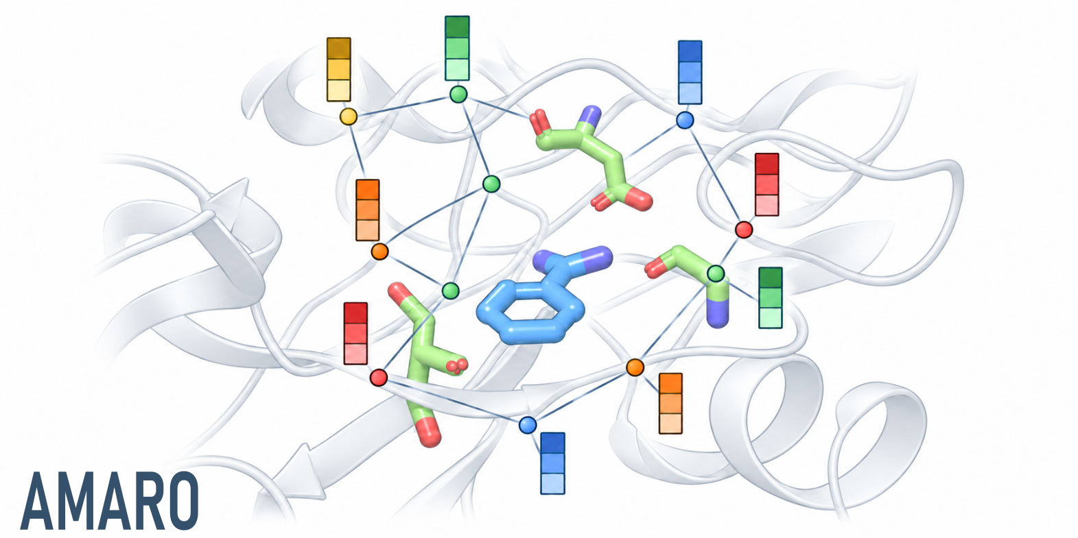
Our new preprint introduces AMARO v2.0, a coarse-grained neural network potential for proteins, drug-like molecules and their complexes, including ternary protein–protein–ligand systems. Developed in collaboration with Universitat Pompeu Fabra (UPF), it brings these molecular classes into one shared model for exploring conformational dynamics and molecular recognition.
One model, with heavy-atom detail
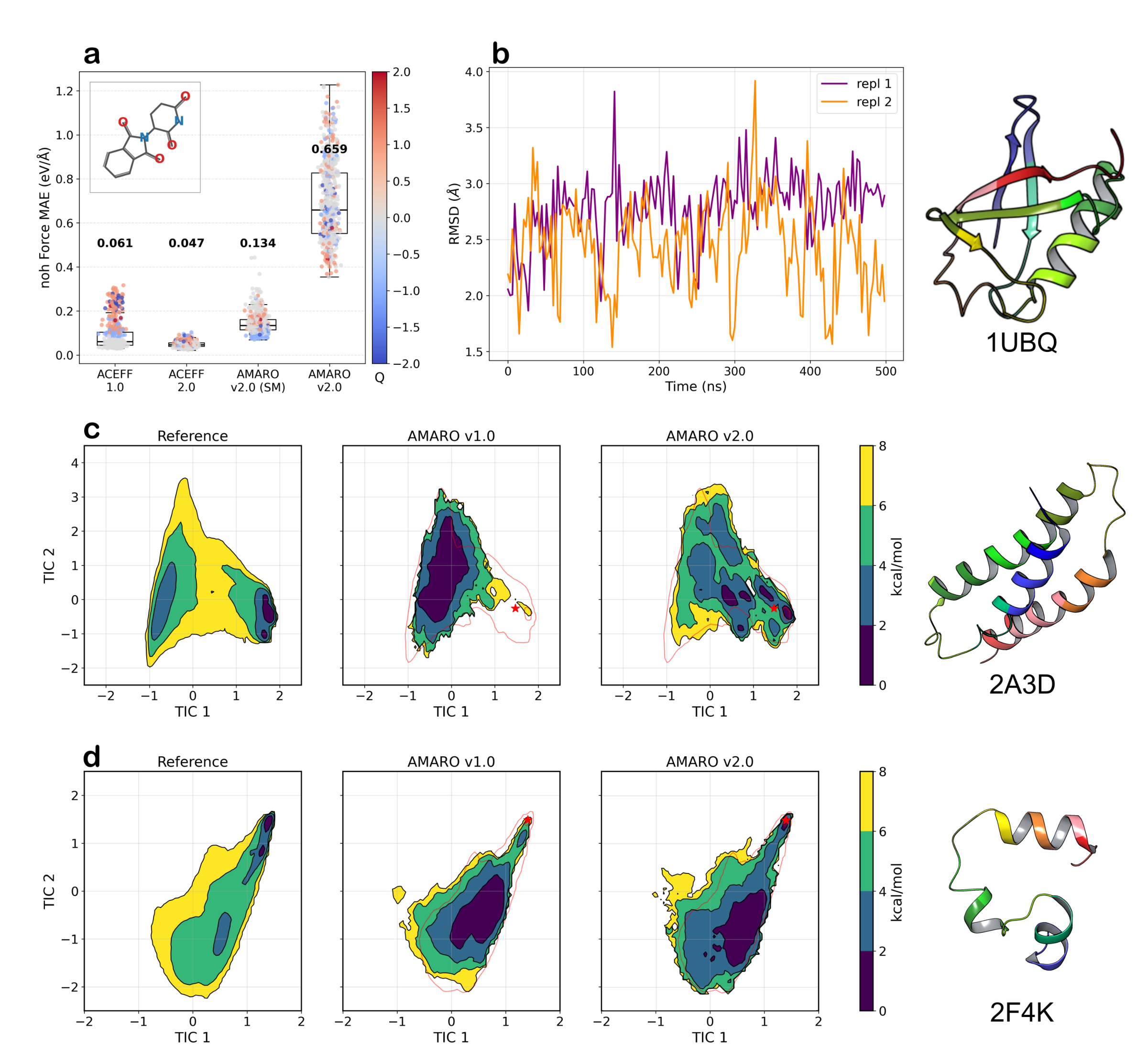
AMARO retains every heavy atom, encodes attached hydrogens in each bead type and conditions the network on total system charge. Solvent is implicit. Its 26 bead types cover 18 elements, extending the original protein model to chemically diverse ligands without separate component potentials. Training proceeds from quantum-mechanical small-molecule forces to protein conformations and then protein–ligand complexes.
Two 500 ns ubiquitin simulations preserve the folded structure. The model also captures key features of the α3D and villin conformational landscapes, improving α3D relative to AMARO v1.0. Adapting the model to complexes does, however, reduce isolated-molecule force accuracy.
More binding-pose recoveries per GPU budget
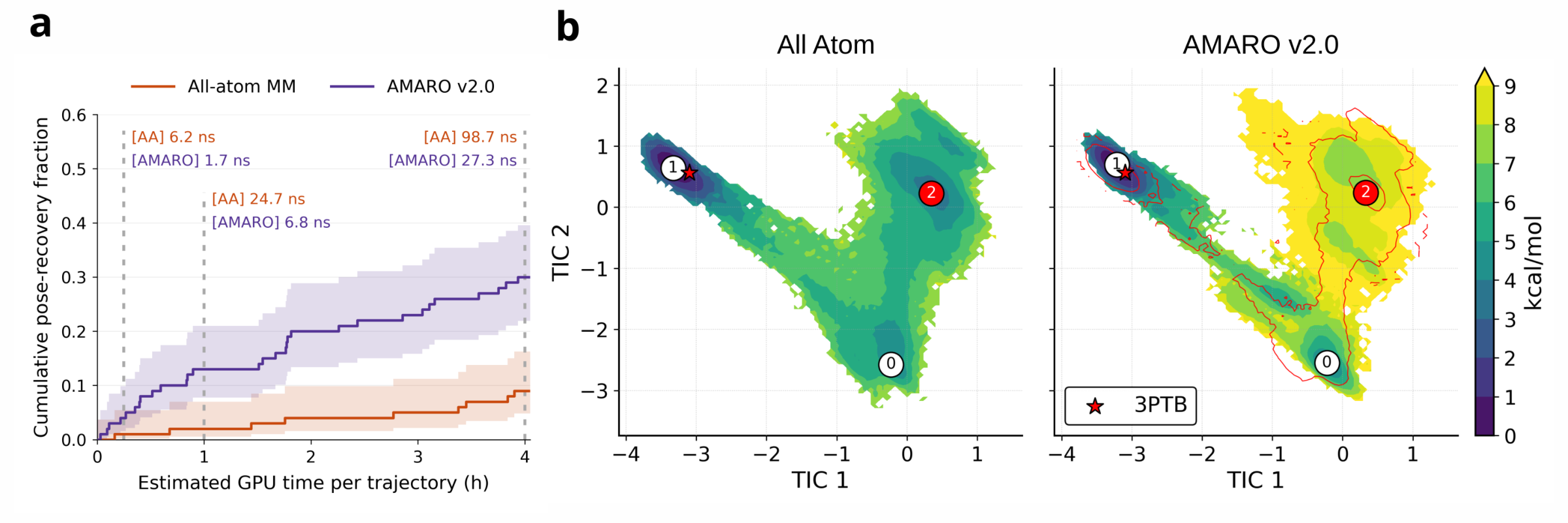
Trypsin–benzamidine tests both chemistry and binding. Protonated benzamidinium maintains stronger contacts with Asp171 and greater pose stability than neutral benzamidine. Starting from unbound configurations, AMARO reaches the crystal pose in 30 of 100 trajectories within an estimated four GPU hours per trajectory, versus 9 of 100 for all-atom molecular mechanics: 3.3-fold more recoveries, despite lower simulated-time throughput. These binding trials use protein Cα restraints and a flat-bottom potential limiting ligand diffusion.
Broader validation, with target-dependent limits
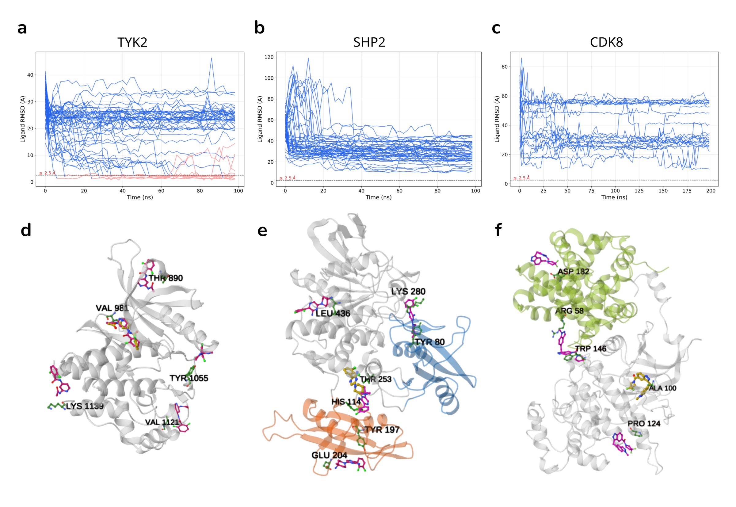
Across 262 complexes spanning 11 targets, most proteins and ligand poses remain close to their starting structures over 10 ns. This is a short-timescale retention test; several receptors closely overlap the training set. In binding trials from unbound configurations, TYK2 recovers the reference pose in 14 of 50 trajectories. SHP2 samples known allosteric regions without recovering the SHP099 pose, while CDK8 predominantly forms off-site contacts.
Binding free-energy estimates are more favorable than experimental references for both trypsin and TYK2, suggesting excessive bound-state stabilization. HIF2A also shows substantial reorientation of its protein partner. These results establish a shared framework for complex dynamics while identifying affinity calibration and interaction specificity as priorities for further development.
Read the preprint: Mirarchi, A.; Farr, S. E.; Perez, A.; De Fabritiis, G. A unified coarse-grained neural network potential for the dynamics of biomolecular complexes. arXiv:2610.37606v1, October 27, 2026. Code and simulation guides on GitHub; model repository on Hugging Face (checkpoints forthcoming).
Molecular glues and PROTACs at Acellera
At Acellera, we will use an enhanced version of this model for molecular glues and PROTACs, where understanding protein–protein–ligand dynamics is central. Write us if you are interested.
AMARO v2.0 in PlayMolecule® AI
AMARO v2.0 is also available through PlayMolecule AI. Acellera’s optimized TensorNet implementation powers these simulations, bringing the shared protein–ligand model into the AI co-scientist interface.